(1) The cause of the disease
The cause of idiopathic Parkinson’s disease is unknown. Some central nervous system degenerative diseases with symptoms of Parkinson disease are mainly degeneration of different parts of the central nervous system, and there are other clinical features, so they can be called symptomatic Parkinson disease, such as progressive supranuclear palsy (PSP), striatal substantia nigra degeneration (SND), Shy-Dräger syndrome (SDS) and olive pontine cerebellar atrophy (OPCA). There are also some diseases or factors that can produce clinical symptoms similar to PD, which are caused by infection, drugs (dopamine blockers, etc.), poisons (MPTP, carbon monoxide, manganese, etc.), vascular (multiple cerebral infarction) and traumatic brain injury, which is clinically called Parkinson’s syndrome (Palkinsonism).
To date, the etiology of PD remains unknown. Current research tends to be related to a combination of factors such as aging, genetic predisposition, and exposure to environmental toxins.
1) Aging: Parkinson’s disease mainly occurs in middle-aged and elderly people, and the onset before the age of 40 is rare, suggesting that aging is related to the onset of the disease. Studies have found that after the age of 30, the activity of dopaminergic neurons, tyrosine oxidase, and dopa decarboxylase in the substantia nigra, and the level of dopamine transmitters in the striatum gradually decrease with age. However, only a small number of elderly people suffer from this disease, indicating that physiological dopaminergic neuronal metamorphosis is not enough to cause the disease, and aging is only a precipitating factor for the onset of this disease.
2) Environmental factors: Epidemiological findings have found regional differences in the prevalence of Parkinson’s disease, so it is suspected that there may be some toxic substances in the environment that damage the neurons of the brain.
3) Genetic predisposition. In recent years, Alα53THr mutations in the common nuclide gene have been found in patients with familial Parkinson’s disease. However, it has not been confirmed many times since.
4) Familial hereditary: In long-term practice, medical scientists have found that Parkinson’s disease seems to have a tendency to cluster in families, and the incidence of Parkinson’s disease patients in their relatives is higher than that of the normal population.
It is generally accepted that Parkinson’s is not a single factor, and that multiple factors may be involved. Genetic factors can increase susceptibility to disease, and only under the interaction with environmental factors and aging, through oxidative stress, mitochondrial failure, calcium overload, excitatory amino acid toxicity, apoptosis, immune abnormalities and other mechanisms, can a large number of dopaminergic neurons in the substantia nigra be degenerated and lost.
(2) Pathogenesis
1. The pathogenesis is very complex and may be related to the following factors.
(1) Aging: PD mainly occurs in middle-aged and elderly people, and the onset before the age of 40 is rare, suggesting that aging is related to the onset of the disease. Studies have found that since the age of 30, the activities of DAergic neurons, tyrosine hydroxylase (TH) and dopa decarboxylase (DDC) in the substantia nigra, the DA transmitter in the striatum have decreased year by year, and the density of DAD1 and D2 receptors has decreased. However, PD is a minority in the elderly, indicating that physiological DAergic neuronal degeneration is not enough to cause the disease. In fact, PD symptoms can only occur clinically if the DAergic neurons in the substantia nigra are reduced by more than 50% and the DA transmitter in the striatum is reduced by more than 80%, and aging is only the precipitating factor of PD.
(2) Environmental factors: Epidemiological investigations have shown that long-term exposure to pesticides, herbicides or certain industrial chemicals may be risk factors for the development of PD. In the early 80s of the 20th century, some drug addicts in California, USA, due to the misuse of a neurotoxic substance, pyridine derivatives 1-methyl4-phenyl 1,2,3,6-tetrahydropyridine (MPTP), showed some pathological changes, biochemical changes, symptoms and drug treatment reactions similar to primary PD, and the injection of MPTP into monkeys also showed similar effects. Neurotoxic MPTP and certain insecticides and herbicides may inhibit the activity of NADH-CoQ reductase (complex I.) in the mitochondrial respiratory chain of the substantia nigra, reduce ATP production and increase free radical production, leading to the degeneration and death of DAergic neurons. There was significant lipid peroxidation and reduced glutathione in the substantia nigra region of PD, suggesting that the antioxidant mechanism disorder and oxidative stress may be related to PD.
(3) Genetic factors: about 10% of patients have a family history, showing autosomal dominant or recessive inheritance with incomplete penetrance, and the rest are sporadic PD. Twin concordance studies have shown that genetic factors may play an important role in some younger (<40 years) patients. So far, 10 single genes such as PARK 1~10 have been identified to be related to PD, and three gene products have been confirmed to be related to familial PD: (1) α-synuclein is a mutation of PARK1 gene, and the gene is located in the long arm of chromosome 4 4q21~23, and α-synuclein may increase the sensitivity of DAergic nerve cells to neurotoxins; (2) Parkin is a mutation of PARK2 gene, which is located in the long arm of chromosome 6 6q25.2~27; (3) Ubiquitin protein C-terminal hydroxylase-L1 is a mutation in the PARK5 gene, located in the short arm of chromosome 4 4p14. Cytochrome P45O2D6 gene and some mitochondrial DNA mutations may be one of the susceptibility factors for PD, which may reduce the activity of P450 enzyme, impaired liver detoxification function, and easily cause damage to the striatum nigra by toxins such as MPTP.
(4) Oxidative stress and free radical generation: Free radicals can cause lipid peroxidation (LPO) of unsaturated fatty acids, which can oxidize and damage proteins and DNA, leading to cell degeneration and death. Due to the increased activity of type B monoamine oxidase (MAO-B), patients with PD can produce excess OH groups and destroy cell membranes. At the same time as oxidation, the DA oxidation products in the substantia nigra cells polymerize to form neuromelanin, which combines with iron to produce a Fenton reaction to form OH. Under normal circumstances, there are enough antioxidant substances in cells, such as glutathione (GSH), glutathione peroxidase (GSH-PX) and superoxide dismutase (SOD) in the brain. In patients with PD, the substantia nigra decreases GSH, increases LPO, increases the concentration of iron ions (Fe2) and decreases ferritin content, making the substantia nigra a site that is susceptible to oxidative stress.
(5) Mitochondrial dysfunction: In recent years, mitochondrial dysfunction has been found to play an important role in the pathogenesis of PD. The understanding of mitochondrial dysfunction in PD patients stems from the study of the mechanism of action of MPTP, which leads to Parkinson disease by inhibiting the activity of mitochondrial respiratory chain complex I in the substantia nigra. In vitro experiments confirmed that MPP, the active ingredient of MPTP, could cause a decrease in mitochondrial membrane potential (ΔΨm) and an increase in oxygen radical production in MES 23.5 cells. The activity of mitochondrial complex I in the substantia nigra in PD patients can be reduced by 32%~38%, and the decrease in the activity of complex α significantly increases the sensitivity of substantia nigra cells to free radical damage. No changes in complex I activity were found in the substantia nigra in patients with multiple system atrophy and progressive supranuclear palsy, suggesting that the decreased activity of PD substantia nigra complex I may be a relative specific change of PD. The presence of mitochondrial dysfunction in PD patients may be related to genetic and environmental factors, and studies have suggested that PD patients have mitochondrial DNA mutations, complex I is encoded and translated by two genomes, nucleus and mitochondria, and any fragment defect of the two sets of genes can affect the function of complex I.
(6) Excitotoxicity: Some authors used microdialysis and HPLC to detect that the content of excitatory amino acids (glutamic acid, aspartic acid) in the striatum of PD monkey model prepared by MPTP was significantly increased. If the concentration of glutamate in the extracellular space is abnormally high, the receptor will be overstimulated, resulting in significant toxic effects on the CNS. Animal experiments have found that intracerebral injection of microscopic glutamate can lead to necrosis of large neurons, glutamate neurotoxicity works through receptors, and NMDA receptor-mediated excitatory neurotoxicity is related to the degeneration of DAergic neurons. Glutamate can damage nerve cells by activating NMDA receptors to produce nitric oxide (NO) and release more excitatory amino acids, further aggravating neuronal damage.
(7) Cytotoxic effect of calcium: human aging can be accompanied by an increase in free Ca2 concentration in nerve cells, a decrease in Ca2/Mg2-ATPase activity, and a decrease in mitochondrial calcium storage capacity. Changes in intracellular Ca2 concentration affect many important functions of neurons, such as cytoskeletal maintenance, neurotransmitter function, protein synthesis and Ca2-mediated enzyme activity, and calbindin, especially 28KD vitamin D-dependent calbindin-D28K, may play an important role, which is related to calcium/magnesium-ATPase activation and has neuroprotective effects. Icopini and Christakos et al. reported that the content of Calbindin-D28K and mRNA expression in the substantia nigra, hippocampus, and dorsal suture nucleus in PD patients were significantly lower than those in normal people, suggesting that the decreased expression of calcium-binding protein gene can also lead to cytotoxicity.
(8) Immunological abnormalities: Abramsky (1978) proposed that the onset of PD is related to immune abnormalities. Clinical studies have found that the cellular immune function of PD patients is reduced, and the activity of interleukin-1 (IL-1) is significantly reduced. McRae-Degueurce et al. reported the presence of anti-DAergic neuronal antibodies in the cerebrospinal fluid (CSF) of PD patients. Cell culture showed that PD plasma and CSF inhibited the function and growth of DAergic neurons in the midbrain of rats. When blood IgG was stereotacticly injected into the substantia nigra on one side of the rat, the substantia nigra tyrosine hydroxylase (TH) and DAergic neurons were significantly reduced, suggesting that it may initiate or participate in immune-mediated substantia nigra cell injury. Tumor necrosis factor-α (TNF-α), IL-6, epithelial growth factor (EGF), metastatic growth factor-α (TGF-α), and β2-microglobulin (β2-MG) may be associated with the pathogenesis of PD.
(9) Apoptosis: Studies have shown that there are apoptosis, free radicals, neurotoxins and neurotrophic factor deficiencies in the pathogenesis of PD. Agid (1995) examined the morphological and biochemical characteristics of apoptosis of DAergic neurons in the substantia nigra of PD patients, and found that about 5-energy neurons in the brain of PD patients had characteristic lesions of apoptosis, and there were TNF-α receptor (α-TN-FR) and bcl-2 proto-oncogene expression, and apoptosis may be the basic step of DAergic neuronal degeneration.
At present, it is generally accepted that PD is not caused by a single factor, and may be involved by multiple factors. Genetic factors increase susceptibility to disease, and under the combined action of environmental factors and aging, the degeneration of DAergic neurons in the substantia nigra is caused by oxidative stress, mitochondrial failure, calcium overload, excitatory amino acid toxicity and apoptosis, resulting in the pathogenesis of the disease.
2. Pathological changes The main lesions of PD are the degeneration and loss of pigment-containing neurons, and the DAergic neurons in the dense part of the substantia nigra are the most significant. Microscopically, nerve cells are reduced, melanogen disappears from substantia nigra cells, and melanin granules are scattered in tissues and macrophages, accompanied by varying degrees of gliosis. Normal human substantia nigra cells decreased with age, substantia nigra cells decreased from 425,000 to 200,000 at the age of 80, and less than 100,000 patients with PD, and more than 50% of DAergic neurons were lost when symptoms appeared, and mild changes were also seen in the locus coeruleus, middle suture nucleus, dorsal vagus nucleus, globus pallidus, putamen nucleus, caudate nucleus and thalamic basil nucleus.
The presence of eosinophilic inclusion bodies in the cytoplasm of residual neurons is an important pathological feature of the disease, and Lewy bodies are glass-like masses composed of cytoplasmic proteins, with a dense core in the center and a filamentous halo around it. A cell can sometimes see multiple Lewy bodies of different sizes, which can be seen in about 10% of the remaining cells, the substantia nigra is obvious, and the globus pallidus, striatum and locus coeruleus are also visible, and α-synuclein and ubiquitin are important components of Lewy bodies.
3. Neurobiochemical changes DA and acetylcholine (Ach), as two important neurotransmitters in the striatum, antagonize each other, and maintaining the balance of the two plays an important role in regulating the activity of the basal ganglia circuit. The DA transmitter pathway in the brain is mainly the substantia nigra-striatal system, and the DAergic neurons in the substantia nigra dense part uptake L-tyrosine from the bloodstream, and form L-dopa (L-dopa) under the action of intracellular tyrosine hydroxylase (TH). Dopamine (DA) is then generated by dopamine decarboxylase (DDC); Through the substantia-striatum nigra-striatum bundle, DA acts on postsynaptic neurons in the putamen nucleus, caudate nucleus, and is finally broken down into homovanillic acid (HVA).
Due to the decrease in TH and DDC in idiopathic Parkinson’s disease, DA production is reduced (L-tyrosine production decreases, and DA production decreases). Monoamine oxidase B (MAO-B) inhibition can reduce the catabolism of DA in neurons and increase the content of DA in the brain. Catechol-oxygen-methyltransferase (COMT) inhibitors can reduce the peripheral metabolism of L-dopa and maintain stable plasma concentrations of L-dopa.
In patients with PD, the loss of DAergic neurons in the substantia nigra, the degeneration of the substantia-striatal DA pathway, and the significant decrease in the DA content of the striatum (>80%), which made the Ach system relatively hyperfunctional, which was the biochemical basis for motor symptoms such as increased muscle tone and decreased movement. In recent years, it has been found that the DA content of the midbrain-limbic system and the midbrain-cortical system is also significantly reduced, which may lead to advanced neurological activity disorders such as mental decline, behavioral and emotional abnormalities, and speech disability. The degree of DA transmitter reduction is consistent with the severity of the patient’s symptoms, and the early stage of the lesion is affected by the increase in DA turnover rate (presynaptic compensation) and the hypersensitivity phenomenon after DA receptor denervation (synaptic postcompensation), the clinical symptoms may be insignificant (compensatory phase), and typical PD symptoms appear with disease progression (decompensated phase). There are also changes in other transmitters or neuropeptides in the basal ganglia, such as norepinephrine (NE), serotonin (5-HT), substance P (SP), enkephalin (ENK), and somatostatin (SS).
Clinical presentation
Parkinson’s disease usually occurs at the age of 40~70, and the incidence increases after the age of 60, and the onset is rare before the age of 30, with only 4 cases in a group of 380 PD patients; Slightly more males. The onset is insidious, the development is slow, and the main manifestations are resting tremor, increased muscle tone and bradykinesia, etc., and the symptoms appear first and then vary from person to person. The first symptom was tremor (60%~70%), followed by walking disorder (12%), muscle rigidity (10%) and bradykinesia (10%). Symptoms often start from one upper limb and gradually spread to the ipsilateral lower limb, contralateral upper limb and lower limb, progressing in an “N” shape (65%~70%); 25%~30% of cases can start from one lower limb, and it is very rare for both lower limbs to start at the same time, and there are still left and right differences in the symptoms of late disease in many cases.
However, regardless of treatment, the chronic progressive course and the need for help after several years are inherent clinical features. A definitive diagnosis can usually be made based on the typical presentation of PD and the positive response to dopa. However, it is difficult to recognize some subclinical symptoms or atypical cases in the early stage, and early diagnosis and early treatment have an important impact on the quality of life in the later stage, which is also the focus of current clinical research. For most patients and clinicians, it is difficult to determine the date of onset of PD, the first symptoms, and the timing of the onset of slowness and tremor. According to the report of Li Danian et al. in China, it is speculated that the preclinical symptoms of PD may be as long as 3~5 years, so the PD symptoms can be divided into two stages: preclinical symptoms and clinical symptoms.
1. Preclinical symptoms The first preclinical symptoms were only reported by Fletcher et al. (1973), but these symptoms have not been taken seriously so far. These symptoms mainly include the following two aspects:
(1) Paresthesias: In fact, as early as Parkinson’s book “Tremor Parsy”, it was described that “some PD cases can have rheumatoid pain before the onset of motor symptoms”, and Charcot also made the same description of 2 PD patients in the same year. It was not until the 70s of the 20th century that Fletcher and Snider et al. gave a more detailed description of the preclinical symptoms and sensory impairment of PD. By the 80s of the 20th century, William et al. combined with electrophysiology to classify sensory impairment, and the sensory symptoms he reported were mainly manifested as unexplained numbness, tingling, ant walking sensation and burning sensation at the joints of the affected limb, mainly at the wrist and ankle, and most of them were intermittent or migratory at first, and fixed in the later stage. There were no obvious objective paresthesias on routine neurological examination, and electrophysiological examination showed somatosensory evoked potentials (SEPs) in some cases, especially the latency and conduction time of the lower limbs. By the early 90s of the 20th century, we conducted a retrospective survey of 150 patients, and the results showed that all patients had experienced paresthesias in the affected limb to varying degrees before the onset of PD clinical symptoms, and this abnormality could persist forever, but it was not parallel to dyspraxia. Electrophysiological examination mainly focuses on somatosensory and cortical evoked potentials, including cortical delay and conduction delay and prolonged latency.
(2) Restless limbs and easy fatigue: In addition to subjective paresthesia, about 1/2 of the patients have experienced indescribable discomfort such as soreness, swelling, numbness or pain in the affected limb in the early stage, and this discomfort mostly occurs or is obvious during rest after exertion, and can be relieved after knocking and beating, which is similar to the manifestation of restless legs syndrome. On the other hand, some patients are prone to fatigue in the affected limbs, especially the wrist and shoulder joints of the upper limbs, and the ankle and knee joints of the lower limbs, and these parts may have slight tremors that are difficult to detect when exerted. General analgesics are effective at first and have no effect after a few months. At this time, the effect of dopa medication can be obvious.
2. Clinical symptoms There were obvious individual differences in the first symptoms, with 85% of the reported subjective paresthesias, 70.5% of tremor, 19.7% of muscle stiffness or slow movements, 12.6% of dexterity and/or dysgraphia, 11.5% of gait disorders, 8.2% of myalgia, cramps and pain, 4.4% of mental disorders such as depression and anxiety and tension, 3.8% of speech disorders, 2.7% of general fatigue or muscle weakness, and 1.6% of drooling and mask face.
(1) Static tremor: It is often the first symptom of PD, and a small number of patients, especially those over 70 years old, can not have tremor. The mechanism is due to the regular, alternating irregular movement of the affected and antagonistic muscles. In the early stages, it usually manifests in the distal extremity, starting on one side, and hand tremor in the upper extremity is more common, and in some patients begins in the knee of the lower extremity. When a rotating component is involved, a ball-like tremor of the thumb and index finger may occur. The tremor frequency is generally 4~8Hz, which appears when stationary, stops when vigorous action, aggravates when nervous, and disappears during sleep. After several years, it affects the ipsilateral upper and lower extremities or the contralateral extremities, and in severe cases, tremors of the head, jaw, lips, tongue, throat, and limbs may occur. Moving one limb, such as clenching or loosening a fist, can cause tremor in the other limb, and this test can help detect early mild tremors. In addition to resting tremor in the later stage, some patients may have action or postural tremor.
(2) Rigidity: Rigidity is one of the main symptoms of PD, mainly due to the increased balance tone of the agonist and antagonist muscles. If it persists in passive motion, it is called “lead tube rigidity or tension”, and if it is accompanied by tremor, a gear-like sensation can be felt during passive movement, which is called “gear-like rigidity or tension”. Myotonia first occurs in the wrist and ankle on the affected side, especially after exertion, and cogwheel muscle tone can be felt when the wrist and ankle are gently and passively moved. Due to the increase in muscle tone, it can bring a series of abnormal symptoms to the patient, such as blinking, chewing, swallowing, walking and other movements.
The following clinical trials are helpful in detecting slight muscle rigidity: (1) muscle rigidity may be more pronounced when the patient is asked to move the contralateral limb; (2) Head dropping test: the patient is in the supine position, and the head often falls slowly when the patient quickly withdraws the pillow under the head, rather than falling quickly; (3) Make the patient put his elbows on the table, so that the forearm is in a perpendicular position to the table, and the muscles of the arms and wrists are relaxed as much as possible, and the wrist joint and the forearm are about 90° flexion at this time in normal people, and the wrist joint of PD patients remains more or less straight, like an erected road sign, which is called the “road sign phenomenon”. Joint pain caused by muscle rigidity in older patients is caused by obstruction of blood supply to the joints due to increased muscle tone.
(3) bradykinesia: decreased voluntary movements, including difficulty initiating and bradykinesia, a series of characteristic dyskinesia symptoms due to increased muscle tone and postural reflex disorder, such as bradykinesia when getting up, turning over, walking and changing direction, decreased facial expression muscle activity, often binocular gaze, reduced blink of an eye, maskedface, difficulty in finger fine movements such as buttoning buttons, tying shoelaces, etc., and the smaller the words become when writing, which is a sign of too little writing ( micrographia) and so on.
Slow or inability to move in patients with PD is a major cause of disability. In the past, it was thought that the movement of PD could not be due to muscle rigidity, but in fact there was no causal relationship between the two. It has been preliminarily demonstrated that decreased motor and inability in PD is a complex symptom that is mainly related to the function of the subcortical extrapyramidal actuator or the impairment of the extrapyramidal descending motor activation device. This is because the symptoms of myotonia are significantly improved after surgery in patients who are unable to exercise, but the frequency of exercise is not consistent with that of dopa.
(4) Abnormal postural gait: postural reflex disorder is the main symptom that brings difficulties to PD patients, and it is second only to decreased movement or akinesia. The patient’s limbs, trunk and neck muscles are stiff and flexed, with the head tilted forward, the trunk bent, the upper limb elbow flexed, the wrist straightened, the forearm adducted, the interphalangeal joint straightened, and the thumb to the palm; The hip and knee joints of the lower limbs are slightly flexed, the lower limbs are dragged in the early stage, and gradually become a small gait, which is difficult to start, rush forward after starting, walk faster and faster, and cannot stop or turn in time, which is called “panic gait” (festination), and the swing of the upper limbs decreases or disappears when walking; When turning, the trunk and head are combined with small steps to turn due to trunk stiffness, which is related to the instability of the center of gravity due to postural balance disorders. Patients are afraid of falling and have to stop when they encounter small obstacles. As the disease progresses, the postural disorder worsens, and it is difficult to stand up in the sitting and lying positions in the late stage. There is currently no clear explanation of the mechanism of this inherent postural reflex disorder in patients with PD, and it has been suggested that this symptom is mainly related to damage to the efferent circuit of the globus pallidus through the thalamus to the cortex.
(5) Other symptoms:
(1) Repeated tapping of the upper edge of the patient’s eyebrow arch can induce non-stop blinking (Myerson sign), and the normal person’s response is not sustained; Blepharoclonus (mild fluttering of closed eyelids) or blepharospasm (involuntary closure of the eyelids) may be present.
(2) Movement disorders of the oral, pharyngeal and palatal muscles, making speech slow, low and monotonous speech, salivation, etc., and difficulty swallowing in severe cases.
(3) It is common to have oily face and excessive sweating caused by hypersecretion of sebaceous glands and sweat glands, intractable constipation caused by gastrointestinal peristalsis disorders, and orthostatic hypotension caused by sympathetic nerve dysfunction, and sphincter function is not affected.
(4) Mental symptoms are more common in depression, anxiety and agitation may occur, and some patients have mild cognitive dysfunction and visual hallucinations in the late stage, which are usually not serious.
3. Clinical classification and classification of PD Written by Wang Xinde, formulated at the National Conference on Extrapyramidal Systems in October 1984.
(1) Primary (idiopathic Parkinson’s disease, i.e., paralysis tremens):
(1) Classification according to the course of the disease:
A. Benign type: the course of the disease is long, with an average of 12 years, and the fluctuation of motor symptoms and the late onset of psychiatric symptoms.
B. Malignant type: the course of the disease is short, up to 4 years on average. Fluctuations in motor symptoms and psychiatric symptoms appear earlier.
(2) Classification according to symptoms:
A. Tremor type.
B. Hypokinetic and tonic type.
C. Hypokinesis tremor and tonic with dementia.
D. Tremor, oligosis and tonic without dementia.
(3) Genetic classification:
A. Familial Parkinson’s disease.
B. Juvenile Parkinson’s disease.
(2) Secondary (Parkinsonism, Symptomatic Parkinsonism):
(1) Infectious (including chronic viral infection) post-encephalitis parkinsonism (narcotic encephalitis, other encephalitis, etc.).
(2) Toxicity (carbon monoxide, manganese, carbon disulfide, hydride, methanol, etc.).
(3) Drug-based (antipsychotic drugs such as phenothiazines, butyrylbenzene, raffle alkaloids and α-methyldopa, etc.).
(4) Cerebrovascular lesions.
(5) Brain tumors (especially midline tumors of the brain).
(6) Traumatic brain injury.
(7) Hollow midbrain.
(8) Metabolic (hypothyroidism, basal ganglia calcification, chronic hepatocerebral degeneration, etc.).
(3) Symptomatic parkinsonism (heterogeneous systemic degeneration):
(1) Progressive supranuclear palsy.
(2) Striatal nigra degeneration.
(3) Cortical dentate nucleus substantia nigra degeneration.
(4) Olivepontine cerebellar atrophy.
(5) Shy-Dräger positional hypotension syndrome.
(6) Dementia [Guam Parkinson-Dementia-Amyotrophic Lateral Sclerosis Syndrome, Jacob-Creutfeldt Disease (Corticostriatal Spinal Degeneration), Alzheimer and Pick Disease, Normal Pressure Hydrocephalus].
(7) Hereditary diseases (Wilson’s disease, Hallerrorden-Speatz disease, Huntington’s disease, spinocerebellar substantia nigra, etc.).
diagnosis
1. Basis for diagnosis
(1) Onset in middle-aged and elderly people, with a slowly progressive course of disease.
(2) At least two of the four main signs (resting tremor, muscle rigidity, bradykinesia, and postural gait disorder), and at least one of the first two signs should be present, and the symptoms should be asymmetrical.
(3) Levodopa treatment is effective, and a positive levodopa test or apomorphine test supports the diagnosis of primary PD.
(4) The patient had no extraocular muscle paralysis, cerebellar signs, orthostatic hypotension, cone system damage and muscle atrophy. The coincidence rate between PD clinical diagnosis and autopsy pathological confirmation was 75%~80%.
2. Commonly used diagnostic and differential diagnostic criteria at home and abroad
(1) Diagnosis of primary Parkinson’s disease (IPD): Wang Xinde wrote the following criteria formulated by the National Conference on Extrapyramidal Systems in October 1984:
(1) At least 2 of the following 4 typical symptoms and signs (resting tremor, oligodimobility, rigidity, positional reflexia) must be present.
and (2) the presence of atypical symptoms and signs that do not support the diagnosis of IPD, such as pyramidal tract signs, apraxia gait disorder, cerebellar symptoms, intention tremor, gaze palsy, severe autonomic dysfunction, and significant dementia with mild extrapyramidal symptoms.
(3) The reduction of homovanillic acid in cerebrospinal fluid is helpful for the diagnosis of early Parkinson’s disease (PD) and essential tremor (ET), drug-induced parkinsonism and PD.
In general, ET is sometimes difficult to distinguish from early IPD, and ET usually presents with positional and action tremors of the hands and head without increased muscle tone and hypomobility.
(2) Diagnosis of secondary parkinsonism (SPDS):
(1) Drug-induced PS (MPS): It is difficult to distinguish drug-induced PS from IPD in clinical practice, and it is important to rely on whether there is a history of taking antipsychotic drugs in the medical history. In addition, the symptoms of drug-induced PS are symmetrical on both sides, and sometimes the symptoms appear first on the side of ADHD. Antipsychotics can be withheld if clinical differentiation is difficult, and PS symptoms usually resolve within a few weeks to 6 months if pharmacologic drugs are present.
(2) Vascular PS (VPS): This sign is characterized by the absence of tremor, often accompanied by focal neurological signs (such as pyramidal tract sign, pseudobulbar palsy, emotional instability, etc.), the course of the disease is mostly step-like, and L-dopa preparation treatment is generally ineffective.
(3) Diagnosis of symptomatic parkinson’s disease syndrome (heterogeneous systemic degeneration):
(1) Progressive supranuclear degeneration: sometimes it is difficult to distinguish from Parkinson’s disease. Progressive supranuclear palsy is clinically characterized by decreased movement, neck rigidity with a later tilt, pseudobulbar palsy, and upward gaze palsy.
(2) Olive pontine cerebellar atrophy: primary Parkinson’s disease should be distinguished from this disease. Olive pontine cerebellar degeneration can also manifest clinically as oligoactivity, rigidity, and even resting tremor. However, cerebellar symptoms such as ataxia are often present at the same time. Characteristic changes may also be seen on CT scanning. Blood glutamate decarboxylase activity is reduced.
(3) Striatal substantia nigra degeneration: This disease and primary Parkinson’s disease are very imaginary, and it is difficult to distinguish them clinically, mainly relying on pathological diagnosis. If L-dopa therapy is clinically ineffective, striatal substantia nigra degeneration should be considered.
(4) Shy-Dräger positional hypotension syndrome: the clinical manifestations are positional hypotension, incontinence, no sweating, and small muscle atrophy of the distal limbs. Sometimes it can be accompanied by Parkinson’s disease. If the patient is clinically found to have parkinson’s disease and symptoms of mild autonomic dysfunction, it needs to be differentiated from primary parkinsonism.
(5) Dementia: It is not uncommon for dementia to be accompanied by Parkinsonism. A. Alzheimer’s disease: In addition to dementia, advanced Alzheimer’s disease has extrapyramidal symptoms, such as oligotransferia, rigidity, and orofacial hyperactivity. In addition, because Parkinson’s disease can be associated with dementia even in the early stages, follow-up is needed to distinguish between the two; B. Normal intracranial pressure hydrocephalus: This disease manifests as gait disorders, urinary incontinence, and dementia. Symptoms of Parkinson’s disease, such as inactivity, rigidity, and resting tremor, can sometimes occur. Computed tomography (CT) is helpful. Radionuclide cisternography is also important in diagnosing normal pressure hydrocephalus.
(6) Hereditary degenerative diseases:
A.苍白球-黑质色素变性病(Hallervorden-Spatz disease)。
B.Huntinton舞蹈病。
C.Lubag(X-连肌张力失常-PDS)。
D. Mitochondrial cell disease with striatal necrosis.
E. Neuroacanthocytosis (β-lipoprotein deficiency).
F. Wilson’s disease (Wilson disease).
Primary PD accounts for 75%~80% of the total number of these clinical types; Secondary (or symptomatic) PD is relatively uncommon; Hereditary degenerative diseases and Parkinson’s superimposed syndrome account for 10%~15%.
IPD is considered in most middle-aged and older patients who have significant slowness, decrease, rigidity, and tremor, while those with early or atypical symptoms are sometimes misdiagnosed. To this end, Takahashi et al. (1992) and Calne et al. (1992) proposed preliminary criteria for the early diagnosis of primary Parkinson’s disease (IPD) and the removal of conditions (Tables 1 and 2).
1. Dietary prescription for Parkinson’s disease (the following information is for reference only, you need to consult a doctor for details)
1. Longan soup with jujube kernels: 15 grams of longan meat and fried jujube kernels. Add longan meat and fried jujube kernels to the water and fry them into juice, and then add an appropriate amount of white honey to serve. Take 2 times daily, morning and evening. It has a tonic effect on people who have suffered from Parkinson’s disease for a long time and qi and blood deficiency.
2. Sea buckthorn chrysanthemum drink: 50 grams of sea buckthorn, 10 grams of chrysanthemum. Wash the sea buckthorn and chrysanthemum and decoction together, 2 times a day, you can take it once in the morning and evening, or you can drink it instead of tea. It is suitable for Parkinson’s disease with hyperlipidemia.
3. Tangerine peel sand kernel sour jujube porridge: 5 grams of tangerine peel, 10 grams of sand kernels, 15 grams of sour jujube, and an appropriate amount of japonica rice. The sand kernels are boiled into soup first, then japonica rice and sour dates are boiled into porridge, and then tangerine peel is added, which can be eaten after a little mixing. Take 2 times a day, morning and evening. Has a sedative effect.
4. Stewed quail with gastrodia
Material: 1 quail, 15 grams of gastrodia.
Production method: Quail hair and internal organs, gastrodia into its belly, tied with thread, stewed with water, add salt and monosodium glutamate.
How to eat: Go to gastrodia, eat meat and drink soup, once every other day.
5. Gastrodia stewed pig brain
Ingredients: 10 grams of gastrodia, 100 grams of pig brain.
Method: Put the above ingredients into a clay pot, add an appropriate amount of water, and simmer for about an hour.
Directions: Drink soup and eat pig brain flower after seasoning, take it once a day, or once every other day.
6. Steamed sheep brain with wolfberry
Ingredients: 50 grams of wolfberry, a sheep brain flower
Production method: Put the above ingredients into a container, add an appropriate amount of water, add minced ginger, green onions, cooking wine, salt, and steam in water before eating.
Directions: Divide into two divided doses daily.
7. Gastrodia fish head soup
Ingredients: 15 grams of gastrodia, 10 grams of Chuanxiong, 1 fresh carp head.
Method: Soak gastrodia and Chuanxiong until soft, cut thin slices into fish heads, put them on a plate, add green onion and ginger, and then add an appropriate amount of water to basket for about 30 minutes.
How to eat: Eat fish and drink soup, once every other day.
8. Goji berry blood vine drink
Ingredients: 20 grams of wolfberry, 15 grams of chicken blood vine, 5 grams of safflower.
Method: Take the above ingredients, add 500 ml of water, fry to 300 ml, pour the liquid medicine into a bowl, and put 30 grams of rice wine.
Directions: Take 1 dose daily in the morning and evening.
9. Walnut yellow wine puree
Ingredients: 15 walnuts, 50 grams of sugar.
Method: Put the above ingredients in a sand pot or porcelain bowl, mash them into a puree with a rolling pin, then put them in a pot, add 50ml of rice wine, and cook over low heat for 10 minutes.
Directions: Take twice daily.
2. What is good for Parkinson’s disease?
(1) General diet: It is basically the same as the diet of normal people, and is suitable for Parkinson’s disease patients with good chewing ability.
(2) Soft food: suitable for patients with reduced chewing ability and digestion, easy to digest (digestible food), easy to chew, soft and non-irritating food.
(3) Semi-liquid soft food: suitable for patients with limited chewing and swallowing functions, you can choose dough sheets, dilute acid, tofu brain, egg custard, egg soup, etc.
(4) Fluid: suitable for patients with advanced disease, obvious impairment of chewing and swallowing function. If you are able to eat by mouth, you should try to eat by mouth and feed slowly with a spoon or bottle to prevent choking. In severe cases, nasogastric feeding is given if necessary. Generally, milk, soybean milk, rice juice, malt milk essence, lotus root flour, broth, vegetable juice, etc. are used as nasogastric diets.
(5) In addition, constipation is common in patients with Parkinson’s disease. It is necessary to give a moderate amount of fresh vegetables (vegetable food), fruits (fruit food) and honey (honey food) in the diet to relieve constipation (constipation food) and supplement vitamins (vitamin food). Avoid irritating foods, tobacco and alcohol.
(6) Eat more cereals, vegetables and fruits. Nutrients such as carbohydrates, protein, dietary fiber, and B vitamins can be obtained from cereals, and the energy required by the body can be obtained. Carbohydrates usually do not affect the efficacy of levodopa.
3. What foods should not be eaten for Parkinson’s disease?
1. Limit protein (protein food). Caloric intake in patients with Parkinson’s disease should be appropriate to maintain normal body weight. Excessive wasting and obesity are not good for treatment. People taking dopamine therapy should limit their protein intake. This is because protein can affect the therapeutic effect of dopamine. Protein intake is limited to less than 0.8 grams per kilogram of body weight per day, and the total amount of protein is about 40 grams ~ 50 grams per day. Within the limit, choose more high-quality proteins such as milk, eggs, meat, and soy products.
2. No smoking, alcohol and irritating foods, such as coffee, chili, mustard, curry, etc.
3. Do not eat fatty meat, meat oil and animal offal, which helps to prevent the adverse effects on the body due to excessive intake of saturated fat and cholesterol. Eating too much fat can also delay the absorption of the levodopa drug, affecting its effectiveness.
1. Prognosis
PD is a chronic progressive disease with no cure, and most patients can continue to work for several years after the onset of the disease, or it can rapidly develop and become disabled. In the later stages of the disease, it can lead to bedridden due to severe muscle rigidity and general stiffness. The cause of death is often complications such as pneumonia and fractures.
2. Recognize the five misunderstandings of Parkinson’s disease:
Myth 1: Smoking can prevent Parkinson’s
Women and children know that smoking is harmful to health (healthy food), but there has been a saying in society that “smoking can prevent Parkinson’s disease”. In fact, this folk claim is not entirely groundless, and studies have now shown that nicotine, the main components of tobacco, as well as 4-benzyridine and hydrazine, can inhibit the toxic effects of some of the neurotoxins that cause Parkinson’s disease, promote their degradation, and increase the level of neurotrophic factors in the brain, thereby protecting dopaminergic neurons in the brain. However, this does not mean that smoking is beneficial to Parkinson’s disease, because at the same time, a large number of investigations and studies have shown that in addition to the increase in the incidence of lung cancer, emphysema and other diseases, smoking has no obvious protective effect on patients with Parkinson’s disease who have been diagnosed, and long-term heavy smoking can lead to cerebral arteriosclerosis, but will increase the chance of suffering from Parkinson’s disease and even other serious diseases such as senile dementia; In addition, many people have the habit of drinking green tea and coffee in their daily lives, and surveys have shown that long-term consumption of green tea and coffee can reduce the incidence of Parkinson’s disease. Current studies have found that tea polyphenols, the main ingredient in green tea, and caffeine, the main ingredient in coffee, can increase the content of dopamine in the brain and inhibit the action of neurotoxins, thereby having a protective effect on dopaminergic neurons, so people who drink it for a long time are not susceptible to Parkinson’s disease. Therefore, maintaining a good lifestyle is very beneficial for the prevention of Parkinson’s disease.
Myth 2: Health (health food) products as a “panacea”
Some patients are often induced by some medical advertisements to mistakenly believe that Parkinson’s disease can be cured with certain home remedies and rashly believe that taking it will not only delay the disease, but also increase the economic burden, and often the gains outweigh the losses. Many people are convinced that ganglioside injections are effective in treating Parkinson’s, but is this the case? Some animal studies have shown that gangliosides can assist in the germination of parabranch nerves, thereby aiding recovery. Hence the theory that gangliosides have a protective effect on cerebral ischemia. But the results of clinical trials in humans don’t really support this idea. As of now, there is no official scientific report, and it has not been clinically proven as a drug for the treatment of Parkinson’s disease! So far this year, 12 randomized controlled trials have been published with 2,265 participants. Most of the trials were of poor quality and their reliability was affected. Disadvantages include vague randomization methods, lack of intention-to-treat analysis, and unclear statistical accounting.
In addition to cerebral infarction, gangliosides are also used to treat spinal cord trauma and brain trauma. Similarly, there is currently a lack of scientific evidence to support this treatment. In the case of spinal cord trauma, initial trials indicated that it might be useful, but at the time it was used in combination with corticosteroids, so efficacy is difficult to determine. Subsequent trials using gangliosides alone have been completed for some time, but detailed results have not yet been published. However, according to the researchers, after six months of medication, there seemed to be no difference between the experimental group and the control group. As for the treatment of brain trauma, there are still no rigorous scientific trials.
Myth 3: Credulous belief that “this knife and that knife” can be cured
The treatment of Parkinson’s disease is usually not solved by one method, and most patients are generally treated with medication, combined with rehabilitation therapy and traditional Chinese medicine. Patients with refractory Parkinson’s disease who are eligible for surgery also require surgical treatment, but they still need to take medication and other treatments after surgery.
After a period of clinical observation, it γ was found that the effect of these treatment methods is not as ideal as imagined, and some can only improve part of the symptoms, and some will even cause serious side effects such as blindness. According to experts, developed countries such as Europe and the United States have basically abandoned these treatments.
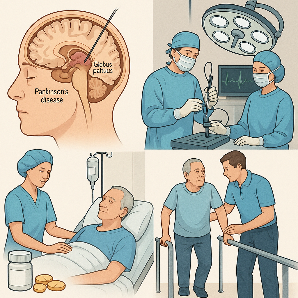
According to reports, at present, the treatment of Parkinson’s disease is recognized as the “gold standard” drug treatment with levodopa, especially compound levodopa, and the other is known as a major breakthrough in the surgical treatment of Parkinson’s disease in recent years, and has been recognized by the medical community as an effective treatment plan for Parkinson’s disease “deep brain electrode stimulation” (commonly known as “brain pacemaker”). In recent years, a new treatment method of “transcranial magnetic stimulation” has been developed abroad, which can be said to fill the gap in physical therapy for Parkinson’s disease.
Myth 4: Mistaken for the treatment of neck and waist diseases
Patients with Parkinson’s disease who have just developed often feel weakness in their limbs, stiffness, stiffness and inflexibility at the ends of their limbs, and sometimes have a feeling of soreness and pain. Doctors also misdiagnose patients with symptoms of one limb in the early stage of the disease as cervical and lumbar spine diseases, and even some patients with Parkinson’s disease with leg onset are mistakenly undergoing intervertebral disc surgery. In fact, the pain sensation of the two diseases is completely different, so please consult neurological and surgical departments if necessary to avoid misdiagnosis.
Myth 5: Hoping for “brain transplant surgery”
At present, some hospitals advertise the so-called “brain transplant surgery”, which is essentially only injected with embryonic brain cell suspension using targeted surgery, and the symptoms are reduced by small local damage, and once the mass effect is removed, the symptoms will reappear. There is also stem cell brain transplantation, which is still in the experimental stage, and although there are cells that survive, they cannot replace the original cells to play a role. In 2003, the US FDA approved an experimental plan to inject a genetically modified virus that can produce dopamine into the brain, which is a new idea and is still in clinical trials. It can be seen that the search for a method that can completely cure Parkinson’s disease needs to be further explored.
Prevention: To date, the exact cause of Parkinson’s disease is not well understood, and as a result, preventive measures are not precisely targeted. However, many studies have confirmed that there is a causal relationship between many of the above-mentioned risk factors and dopamine neuronal degeneration and necrosis in the substantia nigra of the midbrain.
Primary prevention (disease-free prevention)
1. Those with a family history of Parkinson’s disease and related gene carriers, and those who have been exposed to toxic chemicals should be regarded as high-risk groups, and should be closely monitored and followed-up, have regular physical examinations, and strengthen health education and pay attention to self-protection.
2. Intensify the environmental protection of industrial and agricultural production, reduce the discharge of harmful gases, sewage and sewage, and strengthen labor protection for harmful workers.
3. Improve drinking water facilities in rural areas and towns, protect water resources, reduce pollution of river water, reservoir water, pond water and well water, and ensure that the broad masses of the people can drink safe and hygienic drinking water.
4. The elderly should be cautious with phenothiazines, reserpine and butyrylbenzene.
5. Pay attention to the prevention and treatment of geriatric diseases (hypertension, hyperlipidemia, hyperglycemia, cerebral arteriosclerosis, etc.), enhance physical fitness, delay aging, prevent atherosclerosis, and play a certain positive role in the prevention of Parkinson’s disease.
Secondary prevention (early detection, early diagnosis, early treatment)
1. Early diagnosis. The subclinical stage of Parkinson’s disease is long, and if preclinical diagnostic techniques such as olfactory dysfunction, PET scan, mitochondrial DNA, dopamine antibody, cerebrospinal fluid chemistry, electrophysiology and other examinations can be carried out as soon as possible, and the subclinical stage of Parkinson’s disease can be detected as early as possible, and neuroprotective agents (such as vitamin E, SOD, glutathione and glutathione peroxidase, neurotrophic factor, seligrin) may delay the course of the entire clinical period.
2. In the early stage of Parkinson’s disease, although the substantia nigra and striatal nerve cells are reduced, the dopamine secretion is compensatorily increased, and the dopamine content in the brain is not significantly reduced at this time, which is called the compensatory period. However, there are those who advocate early use of low-dose levodopa to reduce complications, which is individualized and preferred. 3. Drug treatment should be used for the decompensated period of Parkinson’s disease.
Tertiary prevention (delay disease progression, prevent disability, improve quality of life)
1. Actively carry out non-drug treatment such as physiotherapy, physical therapy, acupuncture, massage, etc., as well as traditional Chinese and Western medicine or surgery, etc., to delay the development of the disease.
2. Pay attention to psychological counseling and spiritual care, ensure adequate sleep, and avoid emotional tension and excitement, so as to reduce the predisposing factors of aggravation of muscle tremor.
3. Actively encourage patients to take the initiative to exercise, such as eating, dressing, washing, etc. If you have a speech impairment, you can try to practice your pronunciation aloud in front of a mirror. Strengthen joint and muscle strength activities and labor training, maintain limb motor function as much as possible, and pay attention to preventing wrestling and limb deformity and disability.
4. Those who have been bedridden for a long time should strengthen life care, pay attention to cleanliness and hygiene, turn over and pat their backs frequently, and prevent complications such as falling pneumonia and bedsore infection, most of Parkinson’s disease dies from infection of the lungs or other systems such as the urinary system. Pay attention to the nutrition of the diet, give nasogastric feeding if necessary, and keep the bowel and bowel movements smooth. In order to continuously enhance physical fitness, improve immune function and reduce mortality.
Laboratory tests
1. Decreased serum renin activity and tyrosine content; The contents of NE and 5-HT in the substantia nigra and striatum were reduced, and the activity of glutamate decarboxylase (GAD) was reduced by 50% compared with the control group.
2. GABA decreased in CSF, and the content of HVA, the metabolites of DA and 5-HT in CSF, decreased significantly.
3. Biochemical assay Radiotherapy was used to detect the decrease in the content of CSF somatostatin. Urine DA and its metabolites 3-methoxytyramine, 5-HT and epinephrine, NE are also reduced.
Imaging tests
1. CT and MRI imaging manifestations Because Parkinson’s disease is a degenerative disease of the central nervous system, the pathological changes are mainly in the substantia nigra, striatum, globus pallidus, caudate nucleus and cerebral cortex, so CT imaging manifestations, in addition to generalized cerebral atrophy, sometimes basal ganglia calcification. In addition to showing cerebral atrophy such as ventricular enlargement, T2-weighted images often have multiple hyperintense spots in the basal ganglia and white matter.
2. SPECT image performance
(1) Functional imaging through dopamine receptors (DAR): dopamine receptors are widely distributed in dopaminergic pathways in the central nervous system, mainly in the substantia nigra and striatal systems, and DAR (DL) is distributed in the cell bodies of non-cholinergic interneurons in the striatum; DAR (D2) is located in the substantia nigra, striatal dopaminergic neuronal cell body.
SPECT is to diagnose early Parkinson’s disease by injecting radionuclides, currently mainly 123I-IBZM, 131I-IBZM, specific D2 receptor markers, into the human body intravenously through the ratio of radioactivity in the basal ganglia region to the radioactivity of the frontal, occipital or cerebellar lobes, reflecting the number and function of DAR receptors. If patients are treated with dopa preparations early, the onset of contralateral brain DAR (D2) is upregulated. In patients with advanced Parkinson’s disease who have been taking dopa preparations for a long time, the ratio of basal ganglia/occipital lobe and basal ganglia/frontal lobe in the brain is reduced, and SPECT functional imaging can only detect the number of DAR receptors, which can not help confirm whether it is primary Parkinson’s disease, but it can distinguish some secondary Parkinson’s disease, and can also be used as an indicator of the evolution of Parkinson’s disease and the effect of drug treatment.
(2) Functional imaging by dopamine transporter (DAT): It is not clear how dopamine transporter (DAT) transports dopamine (DA), which is mainly distributed in the basal ganglia and thalamus, followed by the frontal lobe. There is a positive correlation between DAT content and the severity of Parkinson’s disease, and the decrease in basal ganglia DAT is significant in patients with early Parkinson’s disease.
SPECT uses 11C-WIN35428 and 123Iβ-CIT to detect the basal ganglia/cerebellar activity ratio and thalamic/cerebellar activity ratio after intravenous injection into the human body, reflecting the number of DAT in different regions of the center. In patients with early Parkinson’s disease, the number of DAT in the basal ganglia region is significantly reduced.
3. PET functional imaging Positron emission tomography (PET) for the diagnosis of Parkinson’s disease, its working principle and method are basically similar to SPECT, at present, it mainly relies on cerebral glucose metabolism scintigraphy, and generally uses 18F deoxyglucose (18FDG). Because in the early stage of Parkinson’s disease, the local glucose metabolism rate of the striatum is moderately reduced, and the glucose metabolism rate is further reduced in the late stage. There are many receptor imaging agents for PET, and PET neurotransmitter functional imaging agents mainly use nuclides such as 18F-dopa-PET (18FD-PET), and the basic principle is the same as SPECT. PET can be used for early diagnosis of Parkinson’s disease, and can be used as an early diagnosis in high-risk groups of Parkinson’s disease, which is an objective indicator to judge the severity of the disease.
Idiopathic PD must be distinguished from familial PD and Parkinson syndrome, and early atypical cases must be distinguished from genetic disorders or degenerative diseases with Parkinson syndrome.
1. Familial PD accounts for about 10%, which is autosomal dominant inheritance of incomplete penetrance, and can be detected by DNA blotting technology, PCR and DNA sequence analysis, etc., to detect α-synuclein gene and Parkin gene mutations, and susceptibility gene analysis such as cytochrome P450-2D6 gene mutations.
2. Parkinson syndrome has a clear cause, secondary to drugs, infection, poisoning, stroke and trauma.
(1) Post-encephalitis Parkinson syndrome: lethargic encephalitis (von Economo) encephalitis that was prevalent in the first half of the 20th century often left parkinsonism, which is rare at present.
(2) Drug or toxic Parkinson syndrome: neuroleptics (phenol-thiazines and butyrylbenzene), reserpine, metoclopramide, α-methyldopa, lithium, flucinazine, etc. can cause Parkinson’s syndrome; Poisoning by MPTP, manganese dust, CO, carbon disulfide or welding fumes can also be caused.
(3) Arteriosclerotic Parkinson syndrome: multiple cerebral infarctions occasionally lead to Parkinson syndrome, patients have a history of hypertension, arteriosclerosis and stroke, and pseudobulbar palsy, pathological signs and neuroimaging examinations can provide evidence.
(4) Traumatic diseases such as boxing encephalopathy, others such as hypothyroidism, hepatocerebral degeneration, brain tumors and normal pressure hydrocephalus can lead to Parkinson syndrome.
3. Genetic disease with Parkinson syndrome
(1) Diffuse Lewis body disease (DLBD): more common in 60~80 years old, dementia, hallucinations, Parkinsonism movement disorders are clinical features, dementia appears early, rapid progression, myoclonus, levodopa response is not good, but the side effects are extremely sensitive.
(2) Wilson’s disease (Wilson disease): it can cause Parkinson’s syndrome, adolescent onset, coarse tremor of one or both upper limbs, muscle rigidity, slow or involuntary movements, liver damage and corneal K-F ring, decreased serum copper, ceruloplasmin, copper oxidase activity, and increased urine copper.
(3) Huntington’s disease: Movement disorders are mainly muscle rigidity and decreased movement, and it is easy to misdiagnose PD.
4. Degenerative disease with Parkinson syndrome
(1) Multiple system atrophy (MSA): involving the basal ganglia, pons, olive, cerebellum and autonomic nervous system, there may be PD-like symptoms, and it is not sensitive to levodopa.
These include: striatal substantia nigra degeneration (SND), which manifests as bradykinesia, muscle rigidity, cone, cerebellum, and autonomic symptoms, and less pronounced tremor. Shy-Dräger syndrome (SDS), prominent autonomic symptoms, orthostatic hypotension, anhidrosis, dysuria, and impotence, as well as pyramidal tract, lower motor neuron, and cerebellar signs. Olive pontine cerebellar atrophy (OPCA), cerebellar and pyramidal system symptoms are prominent, and MRI shows cerebellar and brainstem atrophy.
(2) Progressive supranuclear palsy (PSP): there may be bradykinesia and muscle rigidity, early postural gait instability and falls, inability to gaze vertically, accompanied by frontotemporal dementia, pseudobulbar palsy, dysarthria, pyramidal tract signs, tremor is not obvious, and levodopa response is poor.
(3) Corticobasal degeneration (CBGD): Manifestations of muscle rigidity, bradykinesia, postural instability, dystonia and myoclonus, etc., there may be cortical compound sensory loss, one limb neglect, apraxia, aphasia and dementia and other cortical damage symptoms, eye movement disorders and pathological signs, levodopa treatment is ineffective.
(4) Alzheimer病伴Parkinson综合征。
(5) Depression: there may be poor expression, monotonous speech, and decreased voluntary movements, and PD patients often coexist. Depression lacks muscle rigidity and tremor, and trial treatment with antidepressants may help differentiate.
(6) Essential tremor: early onset, postural or action tremor, affecting the head and causing nodding or shaking, PD typically affects the face and lips. There is no muscle rigidity and bradykinesia in this disease, and about one-third of patients have a family history, and the tremor is significantly reduced by drinking alcohol or taking propranololium.
1. Injury is a complication of Parkinson’s disease that cannot be ignored. As the disease progresses, tremors, stiffness, and coordination dysfunction will gradually affect motor function, and injuries such as fractures may occur when encountering obstacles under the feet. Icy and slippery roads in winter and rainy days, and damp and smooth tiled floors in toilets and bathrooms are dangerous places for people with Parkinson’s disease who are slow and unsteady to walk, so be extra careful to avoid falling.
2. It is often complicated by psychological disorders and mental impairment, especially in advanced patients. Parkinson’s disease manifested by limb tremors, stiffness, clumsy movements, and a mask face due to lack of facial expressions, as well as slurred speech, single intonation, decreased volume, drooling, etc., make patients feel indecent, often have a sense of inferiority complex, unwilling to participate in social activities, do not go to public places, and neglect interpersonal interactions. During treatment and during the development of the disease, insomnia, anxiety, depression, dementia, etc. can also be seen.
3. Autonomic nerve dysfunction leads to the occurrence of digestive system complications. The manifestations are: (1) nutritional disorders and water and electrolyte disorders, which are related to dysphagia, reduced diet, and insufficient fluid supplementation. Dysphagia is caused by a disorder in the coordination of the pharyngeal muscles, which slows the speed of chewing, and as a result, chewing slowly and for longer periods of time, causing food to accumulate in the mouth and throat; Eating too quickly can lead to choking and choking. (2) Esophageal dilation, pseudodiverticulum formation, poor function of esophageal dilator muscle, and burning sensation behind the sternum. Radiologically evidenced of gastric and esophageal reflux. (3) Delayed gastric emptying, some statistics account for about 55%, which is manifested as postprandial fullness, nausea, and vomiting. (4) Poor motor function of the small intestine, resulting in a feeling of abdominal distention. Radiologic studies show small bowel dilation. (5) Poor colon function, mainly manifested as constipation, its high incidence (50%~67%) and stubbornness bring pain to patients, making it difficult for doctors to treat. The various complications of the digestive system have the same pathophysiological basis, which are caused by excessive tension of the gastrointestinal smooth muscles, slow movement, and poor coordination with each other.
4. Infection is a complication that poses a threat to Parkinson’s disease. General respiratory infections and fever can aggravate the symptoms of the disease. Due to low immune function, colds often occur, and patients are also prone to bronchitis, pneumonia, gastroenteritis, etc., and patients who are bedridden in the late stage completely lose their ability to take care of themselves, unable to sit up independently, and even unable to turn over on their own, and malnutrition, skin pressure, often resulting in bedsores. Pneumonitis, aspiration pneumonia, and heart failure are common complications in patients with advanced disease, which can eventually lead to death. Frequent urination is also often the reason for Parkinson’s patients to seek medical attention, especially at night. Men often have an enlarged prostate, which can cause dysuria. Female patients can cause recurrent urinary tract infections until kidney damage due to poor nursing and stool maceration. Infection and sepsis are important causes of death in the advanced stage of the disease.
5. Limb contractures, deformities, joint stiffness, etc. are mainly seen in the late stage of the disease. Therefore, patients in the early and middle stages should be encouraged to exercise more, and patients in the advanced stage should be encouraged to do more passive activities to delay limb complications.
Share this content:
Leave a Reply