Vascular Parkinsonism (VP) often occurs suddenly after an acute stroke or systemic hypoxia, or gradually after multiple strokes. Symptoms are typically asymmetric, and the disease progresses in a stepwise manner.
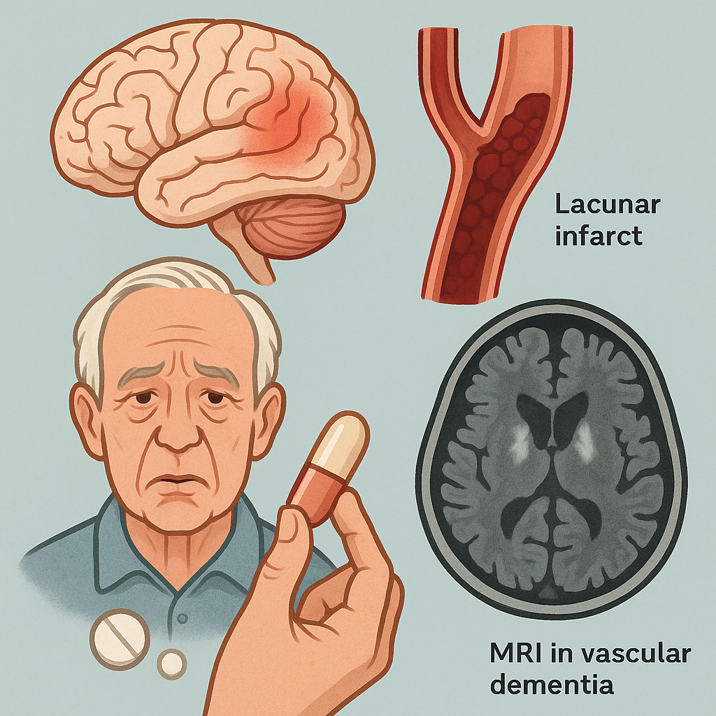
Murrow et al. reported cases with Parkinsonian symptoms and postmortem findings of widespread lacunar infarctions in the basal ganglia (caudate nucleus, internal capsule, globus pallidus, putamen, midbrain), while the substantia nigra remained intact. A Chinese case study confirmed VP via autopsy, revealing bilateral basal ganglia lacunar infarcts and frontal/occipital lobe damage without substantia nigra degeneration. Yamanouchi’s research suggests VP is linked to extensive frontal white matter damage rather than basal ganglia lesions.
Symptoms:
VP primarily manifests as lower-limb motor dysfunction, including a “magnetic gait” (severe difficulty initiating steps), short shuffling steps, pyramidal signs, and dementia. Unlike Parkinson’s disease (PD), VP lacks resting tremors, shows symmetric rigidity, and responds poorly to levodopa. Key distinctions include frequent pyramidal signs, pseudobulbar palsy, and acute/subacute onset.
Diagnosis:
- Clinical Criteria: History of hypertension, diabetes, or stroke; absence of resting tremors; presence of pyramidal signs or dementia.
- Imaging: MRI shows vascular lesions (e.g., lacunar infarcts) in watershed zones, basal ganglia, or frontal white matter. Lesion volume often exceeds 0.6% of total brain tissue.
- Exclusion: Rule out PD, progressive supranuclear palsy (PSP), normal-pressure hydrocephalus, and drug-induced Parkinsonism.
Treatment & Prevention:
Focus on managing cardiovascular risk factors (hypertension, diabetes). Antiplatelet/anticoagulant therapy may reduce VP risk. Levodopa is typically ineffective.
Imaging & Lab Findings:
MRI reveals vascular lesions in subcortical regions. Routine blood tests and CSF analysis are nonspecific.
Differential Diagnoses:
Distinguish from PD, PSP, and normal-pressure hydrocephalus (via CT/MRI showing ventriculomegaly).
Complications:
Linked to stroke, hypertension, atherosclerosis, and cardiovascular diseases.
Keywords with Google Search Links:
- Vascular Parkinsonism
- lacunar infarcts
- basal ganglia lesions
- magnetic gait
- pseudobulbar palsy
- frontal white matter damage
Share this content:
Leave a Reply